Fluorescence microscopy is a microscopy technique, where a certain wavelength of light is shined onto the sample, producing another light wavelength (by fluorescence), which is viewed or measured.
Fluorescence microscopy is very useful for its ability to track a certain molecule or type of molecule dynamically, as well as its high sensitivity to individual molecules. For this reason, it is the tool of choice for most cell and molecular biologists.
In order to harness all the advantages of fluorescence microscopy, we must be able to measure the laser or light source. This is necessary to calibrate the source (in quantitative microscopy) or simply to align the optics properly.
Ok, so let’s Google it: “How to measure laser power”
Here’s the first picture from Google Images:
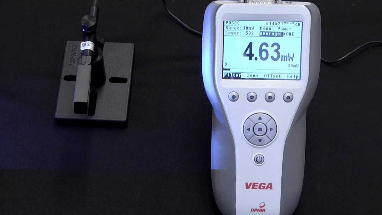
It’s a classic power meter + photodiode sensor setup for measuring low power lasers (up to ~1 W).
So, can we use this to measure our fluorescence microscope?
Perhaps, but it is certainly not ideal. Here’s why:
- Angular dependence – Ophir’s standard photodiode sensors have a high angular dependence, meaning that they will measure less accurately the more on-angle the sensor is placed, relative to the laser beam. Fluorescence microscopy laser sources are usually highly divergent (high N.A.), meaning that wherever the sensor is placed, a large part of the beam will be coming in at a high angle.
- Immersion medium – Fluorescence microscopy optics are often immersed in oil or water, which could damage the sensor or affect its accuracy.
- Physical dimensions – Depending on the microscope, many standard sensors might be awkward or impossible to fit into the path of the laser source for measurement.
If a standard photodiode is not the best way to measure a fluorescence microscope laser source, what is?
PD300-MS Microscope Slide Sensor
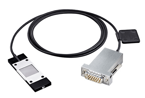
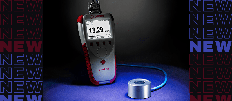
Ophir recently released the PD300-MS sensor designed specifically for measuring fluorescence microscopy light sources.
It was developed to address each of the challenges mentioned above:
-
- Angular dependence – The PD300-MS has a very flat response even at high angles. As you can see in the graph below, it measures within ±3% up to a numerical aperture of about 0.85.

- Immersion medium – The PD300-MS has a smooth cleanable detection area which makes it easy to use with objectives immersed in water or oil. The water or oil can be applied directly to the sensor surface for measurement.
- Physical dependence – The PD300-MS is the size and shape of a microscope slide, so it should fit easily in the fluorescence microscopy – by definition.
As always at Ophir, we are excited and proud to support the cutting edge of science and technology. Whether that means extending to higher and lower powers, new wavelengths, or (as in this case), simply making some design tweaks to support new types of lasers and applications.
You can find out more about the PD300-MS microscope slide sensor here: http://www.ophiropt.com/laser–measurement/laser-power-energy-meters/products/Laser-Photodiode-Sensors/Special-Photodiode-Sensors/PD300-MS
Or contact your local representative:
http://www.ophiropt.com/laser–measurement/contact/power-meters-distributors
- Angular dependence – The PD300-MS has a very flat response even at high angles. As you can see in the graph below, it measures within ±3% up to a numerical aperture of about 0.85.





Leave a Reply
Your email address will not be published. Required fields are marked *